


A medical breakthrough in the treatment of spina bifida has reached a significant milestone, as a world-first clinical trial using in-utero stem-cell therapy has been declared safe. This pioneering approach, which involves treating the condition before a child is even born, marks a transformative moment in foetal medicine. For decades, the medical community has sought ways to mitigate the life-altering effects of myelomeningocele, the most severe form of spina bifida, which often leaves children with permanent paralysis and lifelong complications. The announcement that this novel therapy has passed its initial safety hurdles offers a new glimmer of hope for families facing a prenatal diagnosis that was once synonymous with profound disability.
Spina bifida occurs when the spinal column does not close completely during the early stages of pregnancy, leaving the delicate nerves of the spinal cord exposed to the environment of the womb. This exposure to amniotic fluid can cause progressive and irreversible damage to the neural tissue, leading to a range of physical challenges after birth. While standard foetal surgery, performed while the baby is still in the uterus, has been used for several years to close the spinal opening, it does not always prevent nerve damage.
Many children who undergo standard prenatal surgery still experience significant weakness in their legs or require assistance with bowel and bladder function. The introduction of stem cells into this surgical process represents a shift from simply 'patching' a hole to actively attempting to repair and protect the underlying nervous system.
The safety declaration follows the first phase of a clinical study that monitored a small group of infants who received the treatment. Researchers observed that the combination of traditional surgery and a specialised stem-cell patch did not lead to any adverse complications for either the mothers or the babies. Most importantly, there were no signs of infection, abnormal tissue growth, or tumours at the site of the repair, which are common concerns when introducing cellular therapies. This success has paved the way for more extensive trials, where the focus will shift from safety to the long-term functional benefits for the children involved.
A Breakthrough in Prenatal Intervention
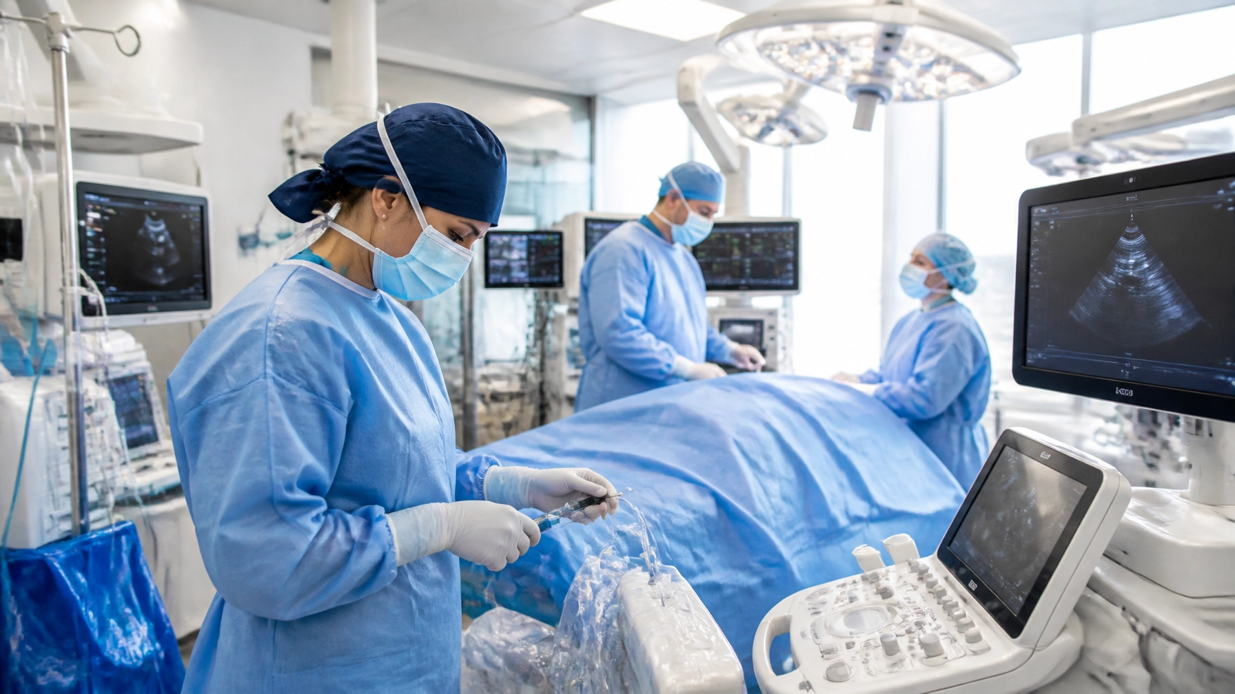
The procedure at the heart of this milestone is known as the CuRe trial, a highly specialised intervention that combines open foetal surgery with a bioengineered stem-cell patch. During the surgery, which is typically performed between the 22nd and 26th weeks of pregnancy, surgeons make an incision in the mother's abdomen and uterus to access the developing foetus. After carefully exposing the spinal defect, the surgical team applies a scaffold seeded with millions of mesenchymal stem cells directly onto the exposed spinal cord. This patch is then secured, and the spinal opening is closed using standard surgical techniques.
The rationale behind this approach is rooted in the "two-hit" hypothesis of spina bifida. The first "hit" is the initial failure of the neural tube to close, and the second "hit" is the ongoing chemical and physical damage caused by the spinal cord's exposure to amniotic fluid. While standard surgery addresses the second hit by providing a protective barrier, it does little to address the damage already sustained or to encourage the regeneration of neural pathways. By placing stem cells directly at the site of the injury, doctors hope to provide a "third hit" of repair, using the cells' natural regenerative properties to salvage nerve function that would otherwise be lost.
The logistical complexity of such a procedure cannot be overstated. It requires a multi-disciplinary team of neurosurgeons, foetal medicine specialists, and stem-cell researchers working in perfect synchrony. The fact that this complex delivery method has been deemed safe is a testament to years of preparation and preclinical research. The surgical team must ensure that the stem cells remain in place despite the fluid environment of the womb and that the mother’s body does not reject the foreign cellular material. The successful births of the first cohort of babies, all of whom reached full term or near-full term without surgical complications related to the patch, confirm that this ambitious protocol is a viable path forward for prenatal care.
The Science of Placental Stem Cells
The specific type of cells used in this therapy are mesenchymal stem cells derived from donated human placentas. These cells were chosen for their unique ability to secrete growth factors and anti-inflammatory signals that can protect damaged nerves. Unlike other types of stem cells that might differentiate into various tissues, these placental cells act more like a "mobile pharmacy," releasing substances that create a healing environment. This helps to reduce the inflammation caused by exposure to amniotic fluid and encourages the survival of existing neurons in the spinal cord.
Before moving to human trials, the effectiveness of these cells was demonstrated in extraordinary preclinical studies. Researchers tested the therapy on lambs that had been induced with spina bifida-like defects. In these animal models, the lambs treated with the stem-cell patch were often able to stand and walk almost normally at birth, whereas those treated with surgery alone remained significantly impaired. These results provided the scientific foundation for the human trial, suggesting that if the therapy worked similarly in humans, it could drastically change the motor outcomes for affected children.
One of the key advantages of using placental-derived cells is their accessibility and their relative safety compared to other sources. Because the placenta is usually discarded after a healthy birth, it provides a sustainable and ethically sound source of high-quality stem cells. Furthermore, these cells appear to have a low risk of causing an immune response, which is crucial for a procedure involving both a mother and her developing child. The focus on safety in the initial phase of the human trial was paramount to ensuring that these cells did not migrate to other parts of the baby’s body or cause any unforeseen developmental issues. To date, the infants monitored in the study have shown no signs of systemic complications, reinforcing the belief that the placenta-derived patch is a safe vehicle for neural repair.
Monitoring Progress and Future Outlook
As the medical community celebrates this safety milestone, the focus now turns to the long-term development of the children who have received the treatment. The first babies treated in the trial are being closely monitored as they reach key developmental milestones, such as sitting up, crawling, and eventually walking. Early observations have been encouraging, with MRI scans showing a reversal of hindbrain herniation in several infants, a common complication of spina bifida where the brain is pulled down towards the spinal column.
This reversal is a positive indicator that the repair is holding and that the intracranial pressure is stabilising.
The children will be followed until they are at least six years old to provide a comprehensive picture of the therapy’s impact. This long-term monitoring is essential for evaluating the most critical measures of success: the ability to walk independently and the management of bowel and bladder function. While it is still too early to declare the treatment a "cure," the absence of significant motor deficits in the early stages of life for some participants is a promising sign. The trial has now entered an expanded phase, allowing more families to participate and providing researchers with a larger data set to determine the treatment's true efficacy compared to standard surgical interventions.
This milestone represents a significant step toward a future where a diagnosis of spina bifida no longer carries the same weight of uncertainty. If the functional benefits observed in animal models translate successfully to the human participants, this stem-cell therapy could become the gold standard for prenatal care. It highlights a broader trend in medicine where the focus is shifting toward regenerative and cellular therapies to treat conditions that were once considered permanent. For the families involved and the medical teams leading the way, the safety declaration is more than just a regulatory hurdle; it is a validation of a decade of research and a bold move toward improving the lives of the next generation. The journey from the laboratory to the operating theatre has been long, but the results so far suggest that the potential for a life without paralysis is closer than ever before.